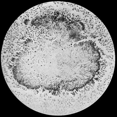
Fig. 30.—Section of Actinomycosis Colony in Pus from Abscess of Liver, showing filaments and clubs of streptothrix actinomyces. × 400 diam. Gram's stain.
Actinomycosis is a chronic disease due to the action of an organism somewhat higher in the vegetable scale than ordinary bacteria—the streptothrix actinomyces or ray fungus.
Fig. 30.—Section of Actinomycosis Colony in Pus from Abscess of Liver, showing filaments and clubs of streptothrix actinomyces. × 400 diam. Gram's stain.
Etiology and Morbid Anatomy.—The actinomyces, which has never been met with outside the body, gives rise in oxen, horses, and other animals to tumour-like masses composed of granulation tissue; and in man to chronic suppurative processes which may result in a condition resembling chronic pyæmia. The actinomyces is more complex in structure than other pathogenic organisms, and occurs in the tissues in the form of small, round, semi-translucent bodies, about the size of a pin-head or less, and consisting of colonies of the fungus. On account of their yellow tint they are spoken of as “sulphur grains.” Each colony is made up of a series of thin, interlacing, and branching filaments, some of which are broken up so as to form masses or chains of cocci; and around the periphery of the colony are elongated, pear-shaped, hyaline, club-like bodies (Fig. 30).
Infection is believed to be conveyed by the husks of cereals, especially barley; and the organism has been found adhering to particles of grain embedded in the tissues of animals suffering from the disease. In the human subject there is often a history of exposure to infection from such sources, and the disease is said to be most common during the harvesting months.
Around each colony of actinomyces is a zone of granulation tissue in which suppuration usually occurs, so that the fungus comes to lie in a bath of greenish-yellow pus. As the process spreads these purulent foci become confluent and form abscess cavities. When metastasis takes place, as it occasionally does, the fungus is transmitted by the blood vessels, as in pyæmia.
Clinical features.—In man the disease may be met with in the skin, the organisms gaining access through an abrasion, and spreading by the formation of new nodules in the same way as tuberculosis.
The region of the mouth and jaws is one of the commonest sites of surgical actinomycosis. Infection takes place, as a rule, along the side of a carious tooth, and spreads to the lower jaw. A swelling is slowly and insidiously developed, but when the loose connective tissue of the neck becomes infiltrated, the spread is more rapid. The whole region becomes infiltrated and swollen, and the skin ultimately gives way and free suppuration occurs, resulting in the formation of sinuses. The characteristic greenish-grey or yellow granules are seen in the pus, and when examined microscopically reveal the colonies of actinomyces.
Less frequently the maxilla becomes affected, and the disease may spread to the base of the skull and brain. The vertebræ may become involved by infection taking place through the pharynx or œsophagus, and leading to a condition simulating tuberculous disease of the spine. When it implicates the intestinal canal and its accessory glands, the lungs, pleura, and bronchial tubes, or the brain, the disease is not amenable to surgical treatment.
Differential Diagnosis.—The conditions likely to be mistaken for surgical actinomycosis are sarcoma, tubercle, and syphilis. In the early stages the differential diagnosis is exceedingly difficult. In many cases it is only possible when suppuration has occurred and the fungus can be demonstrated.
The slow destruction of the affected tissue by suppuration, the absence of pain, tenderness, and redness, simulate tuberculosis, but the absence of glandular involvement helps to distinguish it.
Syphilitic lesions are liable to be mistaken for actinomycosis, all the more that in both diseases improvement follows the administration of iodides. When it affects the lower jaw, in its early stages, actinomycosis may closely simulate a periosteal sarcoma.
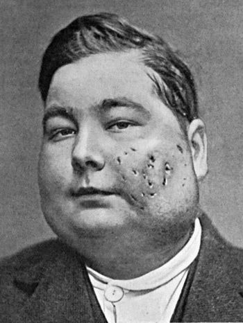
Fig. 31.—Actinomycosis of Maxilla. The disease spread to opposite side; finally implicated base of skull, and proved fatal. Treated by radium.
(Mr. D. P. D. Wilkie's case.)
The recognition of the fungus is the crucial point in diagnosis.
Prognosis.—Spontaneous cure rarely occurs. When the disease implicates internal organs, it is almost always fatal. On external parts the destructive process gradually spreads, and the patient eventually succumbs to superadded septic infection. When, from its situation, the primary focus admits of removal, the prognosis is more favourable.
Treatment.—The surgical treatment is early and free removal of the affected tissues, after which the wound is cauterised by the actual cautery, and sponged over with pure carbolic acid. The cavity is packed with iodoform gauze, no attempt being made to close the wound.
Success has attended the use of a vaccine prepared from cultures of the organism; and the X-rays and radium, combined with the administration of iodides in large doses, or with intra-muscular injections of a 10 per cent. solution of cacodylate of soda, have proved of benefit.
Mycetoma, or Madura Foot.—Mycetoma is a chronic disease due to an organism resembling that of actinomycosis, but not identical with it. It is endemic in certain tropical countries, and is most frequently met with in India. Infection takes place through an abrasion of the skin, and the disease usually occurs on the feet of adult males who work barefooted in the fields.
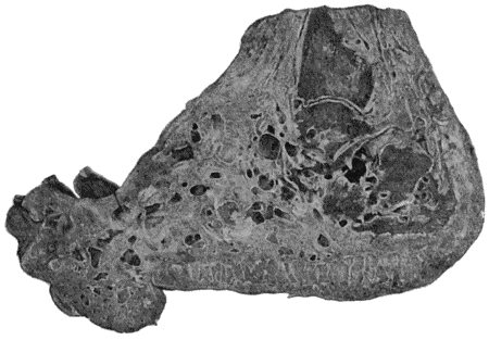
Clinical Features.—The disease begins on the foot as an indurated patch, which becomes discoloured and permeated by black or yellow nodules containing the organism. These nodules break down by suppuration, and numerous minute abscesses lined by granulation tissues are thus formed. In the pus are found yellow particles likened to fish-roe, or black pigmented granules like gunpowder. Sinuses form, and the whole foot becomes greatly swollen and distorted by flattening of the sole and dorsiflexion of the toes. Areas of caries or necrosis occur in the bones, and the disease gradually extends up the leg (Fig. 32). There is but little pain, and no glandular involvement or constitutional disturbance. The disease runs a prolonged course, sometimes lasting for twenty or thirty years. Spontaneous cure never takes place, and the risk to life is that of prolonged suppuration.
If the disease is localised, it may be removed by the knife or sharp spoon, and the part afterwards cauterised. As a rule, amputation well above the disease is the best line of treatment. Unlike actinomycosis, this disease does not appear to be benefited by iodides.
Delhi Boil.—Synonyms—Aleppo boil, Biskra button, Furunculus orientalis, Natal sore.
Delhi boil is a chronic inflammatory disease, most commonly met with in India, especially towards the end of the wet season. The disease occurs oftenest on the face, and is believed to be due to an organism, although this has not been demonstrated. The infection is supposed to be conveyed through water used for washing, or by the bites of insects.
Clinical Features.—A red spot, resembling the mark of a mosquito bite, appears on the affected part, and is attended with itching. After becoming papular and increasing to the size of a pea, desquamation takes place, leaving a dull-red surface, over which in the course of several weeks there develops a series of small yellowish-white spots, from which serum exudes, and, drying, forms a thick scab. Under this scab the skin ulcerates, leaving small oval sores with sharply bevelled edges, and an uneven floor covered with yellow or sanious pus. These sores vary in number from one to forty or fifty. They may last for months and then heal spontaneously, or may continue to spread until arrested by suitable treatment. There is no enlargement of adjacent glands, and but little inflammatory reaction in the surrounding tissues; nor is there any marked constitutional disturbance. Recovery is often followed by cicatricial contraction leading to deformity of the face.
The treatment consists in destroying the original papule by the actual cautery, acid nitrate of mercury, or pure carbolic acid. The ulcers should be scraped with the sharp spoon, and cauterised.
Chigoe.—Chigoe or jigger results from the introduction of the eggs of the sand-flea (Pulex penetrans) into the tissues. It occurs in tropical Africa, South America, and the West Indies. The impregnated female flea remains attached to the part till the eggs mature, when by their irritation they cause localised inflammation with pustules or vesicles on the surface. Children are most commonly attacked, particularly about the toe-nails and on the scrotum. The treatment consists in picking out the insect with a blunt needle, special care being taken not to break it up. The puncture is then cauterised. The application of essential oils to the feet acts as a preventive.
Poisoning by Insects.—The bites of certain insects, such as mosquitoes, midges, different varieties of flies, wasps, and spiders, may be followed by serious complications. The effects are mainly due to the injection of an irritant acid secretion, the exact nature of which has not been ascertained.
The local lesion is a puncture, surrounded by a zone of hyperæmia, wheals, or vesicles, and is associated with burning sensations and itching which usually pass off in a few hours, but may recur at intervals, especially when the patient is warm in bed. Scratching also reproduces the local signs and symptoms. Where the connective tissue is loose—for example, in the eyelid or scrotum—there is often considerable swelling; and in the mouth and fauces this may lead to œdema of the glottis, which may prove fatal.
The treatment consists in the local application of dilute alkalies such as ammonia water, solutions of carbonate or bicarbonate of soda, or sal-volatile. Weak carbolic lotions, or lead and opium lotion, are useful in allaying the local irritation. One of the best means of neutralising the poison is to apply to the sting a drop of a mixture containing equal parts of pure carbolic acid and liquor ammoniæ.
Free stimulation is called for when severe constitutional symptoms are present.
Snake-Bites.—We are here only concerned with the injuries inflicted by the venomous varieties of snakes, the most important of which are the hooded snakes of India, the rattle-snakes of America, the horned snakes of Africa, the viper of Europe, and the adder of the United Kingdom.
While the virulence of these creatures varies widely, they are all capable of producing in a greater or less degree symptoms of acute poisoning in man and other animals. By means of two recurved fangs attached to the upper jaw, and connected by a duct with poison-secreting glands, they introduce into their prey a thick, transparent, yellowish fluid, of acid reaction, probably of the nature of an albumose, and known as the venom.
The clinical features resulting from the injection of the venom vary directly in intensity with the amount of the poison introduced, and the rapidity with which it reaches the circulating blood, being most marked when it immediately enters a large vein. The poison is innocuous when taken into the stomach.
Locally the snake inflicts a double wound, passing vertically into the subcutaneous tissue; the edges of the punctures are ecchymosed, and the adjacent vessels the seat of thrombosis. Immediately there is intense pain, and considerable swelling with congestion, which tends to spread towards the trunk. Extensive gangrene may ensue. There is no special involvement of the lymphatics.
The general symptoms may come on at once if the snake is a particularly venomous one, or not for some hours if less virulent. In the majority of viper or adder bites the constitutional disturbance is slight and transient, if it appears at all. Snake-bites in children are particularly dangerous.
The patient's condition is one of profound shock with faintness, giddiness, dimness of sight, and a feeling of great terror. The pupils dilate, the skin becomes moist with a clammy sweat, and nausea with vomiting, sometimes of blood, ensues. High fever, cramps, loss of sensation, hæmaturia, and melæna are among the other symptoms that may be present. The pulse becomes feeble and rapid, the respiratory nerve centres are profoundly depressed, and delirium followed by coma usually precedes the fatal issue, which may take place in from five to forty-eight hours. If the patient survives for two days the prognosis is favourable.
Treatment.—A broad ligature should be tied tightly round the limb above the seat of infection, to prevent the poison passing into the general circulation, and bleeding from the wound should be encouraged. The application of an elastic bandage from above downward to empty the blood out of the infected portion of the limb has been recommended. The whole of the bite should at once be excised, and crystals of permanganate of potash rubbed into the wound until it is black, or peroxide of hydrogen applied with the object of destroying the poison by oxidation.
The general treatment consists in free stimulation with whisky, brandy, ammonia, digitalis, etc. Hypodermic injections of strychnin in doses sufficiently large to produce a slight degree of poisoning by the drug are particularly useful. The most rational treatment, when it is available, is the use of the antivenin introduced by Fraser and Calmette.