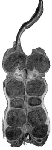
Fig. 66.—Thrombosis in Tortuous and Pouched Great Saphena Vein, in longitudinal section.
The term varix is applied to a condition in which veins are so altered in structure that they remain permanently dilated, and are at the same time lengthened and tortuous. Two types are met with: one in which dilatation of a large superficial vein and its tributaries is the most obvious feature; the other, in which bunches of distended and tortuous vessels develop at one or more points in the course of a vein, a condition to which Virchow applied the term angioma racemosum venosum. The two types may occur in combination.
Any vein in the body may become varicose, but the condition is rare except in the veins of the lower extremity, in the veins of the spermatic cord (varicocele), and in the veins of the anal canal (hæmorrhoids).
We are here concerned with varix as it occurs in the veins of the lower extremity.
Etiology.—Considerable difference of opinion exists as to the essential cause of varix. The weight of evidence is in favour of the view that, when dilatation is the predominant element, it results from a congenital deficiency in the number, size, and strength of the valves of the affected veins, and in an inherent weakness in the vessel walls. The angioma racemosum venosum is probably also due to a congenital alteration in the structure of the vessels, and is allied to tumours of blood vessels. The view that varix is congenital in origin, as was first suggested by Virchow, is supported by the fact that in a large proportion of cases the condition is hereditary; not only may several members of the same family in succeeding generations suffer from varix, but it is often found that the same vein, or segment of a vein, is involved in all of them. The frequent occurrence of varix in youth is also an indication of its congenital origin.
In the majority of cases it is only when some exciting factor comes into operation that the clinical phenomena associated with varix appear. The most common exciting cause is increased pressure within the veins, and this may be produced in a variety of ways. In certain diseases of the heart, lungs, and liver, for example, the venous pressure may be so raised as to cause a localised dilatation of such veins as are congenitally weak. The direct pressure of a tumour, or of the gravid uterus on the large venous trunks in the pelvis, may so obstruct the flow as to distend the veins of the lower extremity. It is a common experience in women that the signs of varix date from an antecedent pregnancy. The importance of the wearing of tight garters as a factor in the production of varicose veins has been exaggerated, although it must be admitted that this practice is calculated to aggravate the condition when it is once established. It has been proved experimentally that the backward pressure in the veins may be greatly increased by straining, a fact which helps to explain the frequency with which varicosity occurs in the lower limbs of athletes and of those whose occupation involves repeated and violent muscular efforts. There is reason to believe, moreover, that a sudden strain may, by rupturing the valves and so rendering them incompetent, induce varicosity independently of any congenital defect. Prolonged standing or walking, by allowing gravity to act on the column of blood in the veins of the lower limbs, is also an important determining factor in the production of varix.
Thrombosis of the deep veins—in the leg, for example—may induce marked dilatation of the superficial veins, by throwing an increased amount of work upon them. This is to be looked upon rather as a compensatory hypertrophy of the superficial vessels than as a true varix.
Morbid Anatomy.—In the lower extremity the varicosity most commonly affects the vessels of the great saphena system; less frequently those of the small saphena system. Sometimes both systems are involved, and large communicating branches may develop between the two.
The essential lesion is the absence or deficiency of valves, so that they are incompetent and fail to support the column of blood which bears back upon them. Normally the valves in the femoral and iliac veins and in the inferior vena cava are imperfectly developed, so that in the erect posture the great saphena receives a large share of the backward pressure of the column of venous blood.
The whole length of the vein may be affected, but as a rule the disease is confined to one or more segments, which are not only dilated, but are also increased in length, so that they become convoluted. The adjacent loops of the convoluted vein are often bound together by fibrous tissue. All the coats are thickened, chiefly by an increased development of connective tissue, and in some cases changes similar to those of arterio-sclerosis occur. The walls of varicose veins are often exceedingly brittle. In some cases the thickening is uniform, and in others it is irregular, so that here and there thin-walled sacs or pouches project from the side of the vein. These pouches vary in size from a bean to a hen's egg, the larger forms being called venous cysts, and being most commonly met with in the region of the saphenous opening and of the opening in the popliteal fascia. Such pouches, being exposed to injury, are frequently the seat of thrombosis (Fig. 66).
Clinical Features.—Varix is most frequently met with between puberty and the age of thirty, and the sexes appear to suffer about equally.
The amount of discomfort bears no direct proportion to the extent of the varicosity. It depends rather upon the degree of pressure in the veins, as is shown by the fact that it is relieved by elevation of the limb. When the whole length of the main trunk of the great saphena is implicated, the pressure in the vein is high and the patient suffers a good deal of pain and discomfort. When, on the contrary, the upper part of the saphena and its valves are intact, and only the more distal veins are involved, the pressure is not so high and there is comparatively little suffering. The usual complaint is of a sense of weight and fulness in the limb after standing or walking, sometimes accompanied by actual pain, from which relief is at once obtained by raising the limb. Cramp-like pains in the muscles are often associated with varix of the deep veins.
The dilated and tortuous vein can be readily seen and felt when the patient is examined in the upright posture. In advanced cases, bead-like swellings are sometimes to be detected over the position of the valves, and, on running the fingers along the course of the vessel, a firm ridge, due to periphlebitis, may be detected on each side of the vein. When the limb is œdematous, the outline of the veins is obscured, but they can be identified on palpation as gutter-like tracks. When large veins are implicated, a distinct impulse on coughing may be seen to pass down as far as the knee; and if the vessel is sharply percussed a fluid wave may be detected passing both up and down the vein.
If the patient is placed on a couch and the limb elevated, the veins are emptied, and if pressure is then made over the region of the saphenous opening and the patient allowed to stand up, so long as the great saphena system alone is involved, the veins fill again very slowly from below. If the small saphena system also is involved, and if communicating branches are dilated, the veins fill up from below more rapidly. When the pressure over the saphenous opening is removed, the blood rapidly rushes into the varicose vessels from above; this is known as Trendelenburg's test.
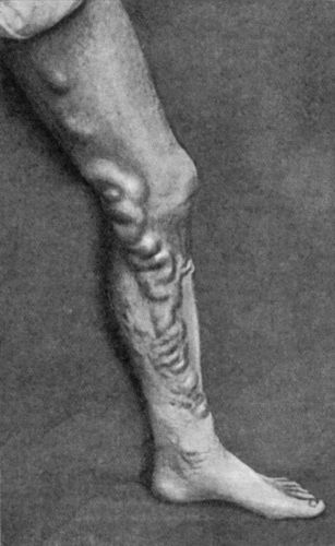
The most marked dilatation usually occurs on the medial side of the limb, between the middle of the thigh and the middle of the calf, the arrangement of the veins showing great variety (Fig. 67).
There are usually one or more bunches of enlarged and tortuous veins in the region of the knee. Frequently a large branch establishes a communication between the systems of the great and small saphenous veins in the region of the popliteal space, or across the front of the upper part of the tibia. The superficial position of this last branch and its proximity to the bone render it liable to injury.
The small veins of the skin of the ankle and foot often show as fine blue streaks arranged in a stellate or arborescent manner, especially in women who have borne children.
Complications.—When the varix is of long standing, the skin in the lower part of the leg sometimes assumes a mahogany-brown or bluish hue, as a result of the deposit of blood pigment in the tissues, and this is frequently a precursor of ulceration.
Chronic dermatitis (varicose eczema) is often met with in the lower part of the leg, and is due to interference with the nutrition of the skin. The incompetence of the valves allows the pressure in the varicose veins to equal that in the arterioles, so that the capillary circulation is impeded. From the same cause the blood in the deep veins is enabled to enter the superficial veins, where the backward pressure is so great that the blood flows down again, and so a vicious circle is established. The blood therefore loses more and more of its oxygen, and so fails to nourish the tissues.
The ulcer of the leg associated with varicose veins has already been described.
Hæmorrhage may take place from a varicose vein as a result of a wound or of ulceration of its wall. Increased intra-venous pressure produced by severe muscular strain may determine rupture of a vein exposed in the floor of an ulcer. If the limb is dependent, the incompetency of the valves permits of rapid and copious bleeding, which may prove fatal, particularly if the patient is intoxicated when the rupture takes place and no means are taken to arrest the hæmorrhage. The bleeding may be arrested at once by elevating the limb, or by applying pressure directly over the bleeding point.
Phlebitis and thrombosis are common sequelæ of varix, and may prove dangerous, either by spreading into the large venous trunks or by giving rise to emboli. The larger the varix the greater is the tendency for a thrombus to spread upwards and to involve the deep veins. Thrombi usually originate in venous cysts or pouches, and at acute bends on the vessel, especially when these are situated in the vicinity of the knee, and are subjected to repeated injuries—for example in riding. Phleboliths sometimes form in such pouches, and may be recognised in a radiogram. In a certain proportion of cases, especially in elderly people, the occurrence of thrombosis leads to cure of the condition by the thrombus becoming organised and obliterating the vein.
Treatment.—At best the treatment of varicose veins is only palliative, as it is obviously impossible to restore to the vessels their normal structure. The patient must avoid wearing anything, such as a garter, which constricts the limb, and any obvious cause of direct pressure on the pelvic veins, such as a tumour, persistent constipation, or an ill-fitting truss, should be removed. Cardiac, renal, or pulmonary causes of venous congestion must also be treated, and the functions of the liver regulated. Severe forms of muscular exertion and prolonged standing or walking are to be avoided, and the patient may with benefit rest the limb in an elevated position for a few hours each day. To support the distended vessels, a closely woven silk or worsted stocking, or a light and porous form of elastic bandage, applied as a puttee, should be worn. These appliances should be put on before the patient leaves his bed in the morning, and should only be removed after he lies down at night. In this way the vessels are never allowed to become dilated. Elastic stockings, and bandages made entirely of india-rubber, are to be avoided. In early and mild cases these measures are usually sufficient to relieve the patient's discomfort.
Operative Treatment.—In aggravated cases, when the patient is suffering pain, when his occupation is interfered with by repeated attacks of phlebitis, or when there are large pouches on the veins, operative treatment is called for. The younger the patient the clearer is the indication to operate. It may be necessary to operate to enable a patient to enter one of the public services, even although no symptoms are present. The presence of an ulcer does not contra-indicate operation; the ulcer should be excised, and the raw surface covered with skin grafts, before dealing with the veins.
The operation of Trendelenburg is especially appropriate to cases in which the trunk of the great saphena vein in the thigh is alone involved. It consists in exposing three or four inches of the vein in its upper part, applying a ligature at the upper and lower ends of the exposed portion, and, after tying all tributary branches, resecting this portion of the vein.
The procedure of C. H. Mayo is adapted to cases in which it is desirable to remove longer segments of the veins. It consists in the employment of special instruments known as “ring-enucleators” or “vein-strippers,” by means of which long portions of the vein are removed through comparatively small incisions.
An alternative procedure consists in avulsing segments of the vein by means of Babcock's stylet, which consists of a flexible steel rod, 30 inches in length, with acorn-shaped terminals. The instrument is passed along the lumen of the segment to be dealt with, and a ligature applied around the vein above the bulbous end of the stylet enables nearly the whole length of the great saphena vein to be dragged out in one piece. These methods are not suitable when the veins are brittle, when there are pouches or calcareous deposits in their walls, or where there has been periphlebitis binding the coils together.
Mitchell of Belfast advises exposing the varices at numerous points by half-inch incisions, and, after clamping the vein between two pairs of forceps, cutting it across and twisting out the segments of the vein between adjacent incisions. The edges of the incisions are sutured; and the limb is firmly bandaged from below upwards, and kept in an elevated position. We have employed this method with satisfactory results.
The treatment of the complications of varix has already been considered.