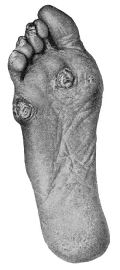
Fig. 93.—Callosities and Corns on the Sole and Plantar Aspect of the Toes in a woman who was also the subject of flat-foot.
Structure of Skin.—The skin is composed of a superficial cellular layer—the epidermis, and the corium or true skin. The epidermis is differentiated from without inwards into the stratum corneum, the stratum lucidum, the stratum granulosum, and the rete Malpighii or germinal layer, from which all the others are developed. The corium or true skin consists of connective tissue, in which ramify the blood vessels, lymphatics, and nerves. That part of the corium immediately adjoining the epidermis is known as the papillary portion, and contains the terminal loops of the cutaneous blood vessels and the terminations of the cutaneous nerves. The deeper portion of the true skin is known as the reticular portion, and is largely composed of adipose tissue.
Blisters result from the exudation of serous fluid beneath the horny layer of the epidermis. The fluid may be clear, as in the blisters of a recent burn, or blood-stained, as in the blisters commonly accompanying fractures of the leg. It may become purulent as a result of infection, and this may be the starting-point of lymphangitis or cellulitis.
The skin should be disinfected and the blisters punctured. When infected, the separated horny layer must be cut away with scissors to allow of the necessary purification.
Fig. 93.—Callosities and Corns on the Sole and Plantar Aspect of the Toes in a woman who was also the subject of flat-foot.
Callosities are prominent, indurated masses of the horny layer of the epidermis, where it has been exposed to prolonged friction and pressure. They occur on the fingers and hand as a result of certain occupations and sports, but are most common under the balls of the toes or heel. A bursa may form beneath a callosity, and if it becomes inflamed may cause considerable suffering; if suppuration ensues, a sinus may form, resembling a perforating ulcer of the foot.
The treatment of callosities on the foot consists in removing pressure by wearing properly fitting boots, and in applying a ring pad around the callosity; another method is to fit a sock of spongiopilene with a hole cut out opposite the callosity. After soaking in hot water, the overgrown horny layer is pared away, and the part painted daily with a saturated solution of salicylic acid in flexile collodion.
Corns.—A corn is a localised overgrowth of the horny layer of the epidermis, which grows downwards, pressing upon and displacing the sensitive papillæ of the corium. Corns are due to the friction and pressure of ill-fitting boots, and are met with chiefly on the toes and sole of the foot. A corn is usually hard, dry, and white; but it may be sodden from moisture, as in “soft corns” between the toes. A bursa may form beneath a corn, and if inflamed constitutes one form of bunion. When suppuration takes place in relation to a corn, there is great pain and disability, and it may prove the starting-point of lymphangitis.
The treatment consists in the wearing of properly fitting boots and stockings, and, if the symptoms persist, the corn should be removed. This is done after the manner of chiropodists by digging out the corn with a suitably shaped knife. A more radical procedure is to excise, under local anæsthesia, the portion of skin containing the corn and the underlying bursa. The majority of so-called corn solvents consist of a solution of salicylic acid in collodion; if this is painted on daily, the epidermis dies and can then be pared away. The unskilful paring of corns may determine the occurrence of senile gangrene in those who are predisposed to it by disease of the arteries.
Chilblains.—Chilblain or erythema pernio is a vascular disturbance resulting from the alternate action of cold and heat on the distal parts of the body. Chilblains are met with chiefly on the fingers and toes in children and anæmic girls. In the mild form there is a sensation of burning and itching, the part becomes swollen, of a dusky red colour, and the skin is tense and shiny. In more severe cases the burning and itching are attended with pain, and the skin becomes of a violet or wine-red colour. There is a third degree, closely approaching frost-bite, in which the skin tends to blister and give way, leaving an indolent raw surface popularly known as a “broken chilblain.”
Those liable to chilblains should take open-air exercise, nourishing food, cod-liver oil, and tonics. Woollen stockings and gloves should be worn in cold weather, and sudden changes of temperature avoided. The symptoms may be relieved by ichthyol ointment, glycerin and belladonna, or a mixture of Venice turpentine, castor oil, and collodion applied on lint which is wrapped round the toe. Another favourite application is one of equal parts of tincture of capsicum and compound liniment of camphor, painted over the area night and morning. Balsam of Peru or resin ointment spread on gauze should be applied to broken chilblains. The most effective treatment is Bier's bandage applied for about six hours twice daily; it can be worn while the patient is following his occupation; in chronic cases this may be supplemented with hot-air baths.
Boils and Carbuncles.—These result from infection with the staphylococcus aureus, which enters the orifices of the ducts of the skin under the influence of friction and pressure, as was demonstrated by the well-known experiment of Garrè, who produced a crop of pustules and boils on his own forearm by rubbing in a culture of the staphylococcus aureus.
A boil results when the infection is located in a hair follicle or sebaceous gland. A hard, painful, conical swelling develops, to which, so long as the skin retains its normal appearance, the term “blind boil” is applied. Usually, however, the skin becomes red, and after a time breaks, giving exit to a drop or two of thick pus. After an interval of from six to ten days a soft white slough is discharged; this is known as the “core,” and consists of the necrosed hair follicle or sebaceous gland. After the separation of the core the boil heals rapidly, leaving a small depressed scar.
Boils are most frequently met with on the back of the neck and the buttocks, and on other parts where the skin is coarse and thick and is exposed to friction and pressure. The occurrence of a number or a succession of boils is due to spread of the infection, the cocci from the original boil obtaining access to adjacent hair follicles. The spread of boils may be unwittingly promoted by the use of a domestic poultice or the wearing of infected underclothing.
While boils are frequently met with in debilitated persons, and particularly in those suffering from diabetes or Bright's disease, they also occur in those who enjoy vigorous health. They seldom prove dangerous to life except in diabetic subjects, but when they occur on the face there is a risk of lymphatic and of general pyogenic infection. Boils may be differentiated from syphilitic lesions of the skin by their acute onset and progress, and by the absence of other evidence of syphilis; and from the malignant or anthrax pustule by the absence of the central black eschar and of the circumstances which attend upon anthrax infection.
Treatment.—The skin of the affected area should be painted with iodine, and a Klapp's suction bell applied thrice daily. If pus forms, the skin is frozen with ethyl-chloride and a small incision made, after which the application of the suction bell is persevered with. The further treatment consists in the use of diluted boracic or resin ointment. In multiple boils on the trunk and limbs, lysol or boracic baths are of service; the underclothing should be frequently changed, and that which is discarded must be disinfected. In patients with recurrence of boils about the neck, re-infection frequently takes place from the scalp, to which therefore treatment should be directed.
Any impaired condition of health should be corrected; when, there is sugar or albumen in the urine the conditions on which these depend must receive appropriate treatment. When there are successive crops of boils, recourse should be had to vaccines. In refractory cases benefit has followed the subcutaneous injection of lipoid solution containing tin.
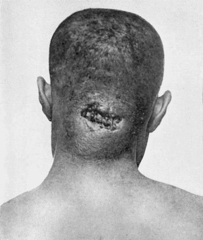
Carbuncle may be looked upon as an aggregation of boils, and is characterised by a densely hard base and a brownish-red discoloration of the skin. It is usually about the size of a crown-piece, but it may continue to enlarge until it attains the size of a dinner-plate. The patient is ill and feverish, and the pain may be so severe as to prevent sleep. As time goes on several points of suppuration appear, and when these burst there are formed a number of openings in the skin, giving it a cribriform appearance; these openings exude pus. The different openings ultimately fuse and the large adherent greyish-white slough is exposed. The separation of the slough is a tedious process, and the patient may become exhausted by pain, discharge, and toxin absorption. When the slough is finally thrown off, a deep gap is left, which takes a long time to heal. A large carbuncle is a grave disease, especially in a weakly person suffering from diabetes or chronic alcoholism; we have on several occasions seen diabetic coma supervene and the patient die without recovering consciousness. In the majority of cases the patient is laid aside for several months. It is most common in male adults over forty years of age, and is usually situated on the back between the shoulders. When it occurs on the face or anterior part of the neck it is especially dangerous, because of the greater risk of dissemination of the infection.
A carbuncle is to be differentiated from an ulcerated gumma and from anthrax pustule.
Treatment.—Pain is relieved by full doses of opium or codein, and these drugs are specially indicated when sugar is present in the urine. Vaccines may be given a trial. The diet should be liberal and easily digested, and strychnin and other stimulants may be of service. Locally the treatment is carried out on the same lines as for boils.
In some cases it is advisable to excise the carbuncle or to make incisions across it in different directions, so that the resulting wound presents a stellate appearance.
Acute Abscesses of the Skin and Subcutaneous Tissue in Young Children.—In young infants, abscesses are not infrequently met with scattered over the trunk and limbs, and are probably the result of infection of the sebaceous glands from dirty underclothing. The abscesses should be opened, and the further spread of infection prevented by cleansing of the skin and by the use of clean under-linen. Similar abscesses are met with on the scalp in association with eczema, impetigo, and pediculosis.
Veldt Sore.—This sore usually originates in an abrasion of the epidermis, such as a sun blister, the bite of an insect, or a scratch. A pustule forms and bursts, and a brownish-yellow scab forms over it. When this is removed, an ulcer is left which has little tendency to heal. These sores are most common about the hands, arms, neck, and feet, and are most apt to occur in those who have had no opportunities of washing, and who have lived for a long time on tinned foods.
Tuberculosis of the Skin.—Interest attaches chiefly to the primary forms of tuberculosis of the skin in which the bacilli penetrate from without—inoculation tubercle and lupus.
Inoculation Tubercle.—The appearances vary with the conditions under which the inoculation takes place. As observed on the fingers of adults, the affection takes the form of an indolent painless swelling, the epidermis being red and glazed, or warty, and irregularly fissured. Sometimes the epidermis gives way, forming an ulcer with flabby granulations. The infection rarely spreads to the lymphatics, but we have seen inoculation tubercle of the index-finger followed by a large cold abscess on the median side of the upper arm and by a huge mass of breaking down glands in the axilla.
In children who run about barefooted in towns, tubercle may be inoculated into wounds in the sole or about the toes, and although the local appearances may not be characteristic, the nature of the infection is revealed by its tendency to spread up the limb along the lymph vessels, giving rise to abscesses and fungating ulcers in relation to the femoral glands.
Tuberculous Lupus.—This is an extremely chronic affection of the skin. It rarely extends to the lymph glands, and of all tuberculous lesions is the least dangerous to life. The commonest form of lupus—lupus vulgaris—usually commences in childhood or youth, and is most often met with on the nose or cheek. The early and typical appearance is that of brownish-yellow or pink nodules in the skin, about the size of hemp seed. Healing frequently occurs in the centre of the affected area while the disease continues to extend at the margin.
When there is actual destruction of tissue and ulceration—the so-called “lupus excedens” or “ulcerans”—healing is attended with cicatricial contraction, which may cause unsightly deformity. When the cheek is affected, the lower eyelid may be drawn down and everted; when the lips are affected, the mouth may be distorted or seriously diminished in size. When the nose is attacked, both the skin and mucous surfaces are usually involved, and the nasal orifices may be narrowed or even obliterated; sometimes the soft parts, including the cartilages, are destroyed, leaving only the bones covered by tightly stretched scar tissue.
The disease progresses slowly, healing in some places and spreading at others. The patient complains of a burning sensation, but little of pain, and is chiefly concerned about the disfigurement. Nothing is more characteristic of lupus than the appearance of fresh nodules in parts which have already healed. In the course of years large tracts of the face and neck may become affected. From the lips it may spread to the gum and palate, giving to the mucous membrane the appearance of a raised, bright-red, papillary or villous surface. When the disease affects the gums, the teeth may become loose and fall out.
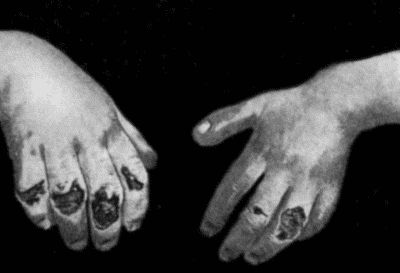
On parts of the body other than the face, the disease is even more chronic, and is often attended with a considerable production of dense fibrous tissue—the so-called fibroid lupus. Sometimes there is a warty thickening of the epidermis—lupus verrucosus. In the fingers and toes it may lead to a progressive destruction of tissue like that observed in leprosy, and from the resulting loss of portions of the digits it has been called lupus mutilans. In the lower extremity a remarkable form of the disease is sometimes met with, to which the term lupus elephantiasis (Fig. 96) has been applied. It commences as an ordinary lupus of the toes or dorsum of the foot, from which the tuberculous infection spreads to the lymph vessels, and the limb as a whole becomes enormously swollen and unshapely.
Finally, a long-standing lupus, especially on the cheek, may become the seat of epithelioma—lupus epithelioma—usually of the exuberant or cauliflower type, which, like other epitheliomas that originate in scar tissue, presents little tendency to infect the lymphatics.
The diagnosis of lupus is founded on the chronic progress and long duration, and the central scarring with peripheral extension of the disease. On the face it is most liable to be confused with syphilis and with rodent cancer. The syphilitic lesion belongs to the tertiary period, and although presenting a superficial resemblance to tuberculosis, its progress is more rapid, so that within a few months it may involve an area of skin as wide as would be affected by lupus in as many years. Further, it readily yields to anti-syphilitic treatment. In cases of tertiary syphilis in which the nose is destroyed, it will be noticed that the bones have suffered most, while in lupus the destruction of tissue involves chiefly the soft parts.
Rodent cancer is liable to be mistaken for lupus, because it affects the same parts of the face; it is equally chronic, and may partly heal. It begins later in life, however, the margin of the ulcer is more sharply defined, and often presents a “rolled” appearance.
Treatment.—When the disease is confined to a limited area, the most rapid and certain cure is obtained by excision; larger areas are scraped with the sharp spoon. The ray treatment includes the use of luminous, Röntgen, or radium rays, and possesses the advantage of being comparatively painless and of being followed by the least amount of scarring and deformity.
Encouraging results have also been obtained by the application of carbon dioxide snow.
Multiple subcutaneous tuberculous nodules are met with chiefly in children. They are indolent and painless, and rarely attract attention until they break down and form abscesses, which are usually about the size of a cherry, and when these burst sinuses or ulcers result. If the overlying skin is still intact, the best treatment is excision. If the abscess has already infected the skin, each focus should be scraped and packed.
Sporotrichosis is a mycotic infection due to the sporothrix Shenkii. It presents so many features resembling syphilis and tubercle that it is frequently mistaken for one or other of these affections. It occurs chiefly in males between fifteen and forty-five, who are farmers, fruit and vegetable dealers, or florists. There is usually a history of trauma of the nature of a scratch or a cut, and after a long incubation period there develop a series of small, hard, round nodules in the skin and subcutaneous tissue which, without pain or temperature, soften into cold abscesses and leave indolent ulcers or sinuses. The infection is of slow progress and follows the course of the lymphatics. From the gelatinous pus the organism is cultivated without difficulty, and this is the essential step in arriving at a diagnosis. The disease yields in a few weeks to full doses of iodide of potassium.
Elephantiasis.—This term is applied to an excessive enlargement of a part depending upon an overgrowth of the skin and subcutaneous cellular tissue, and it may result from a number of causes, acting independently or in combination. The condition is observed chiefly in the extremities and in the external organs of generation.
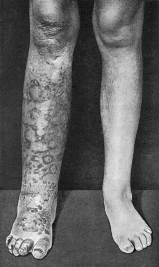
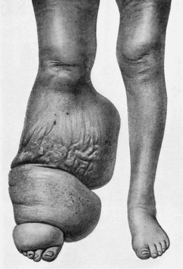
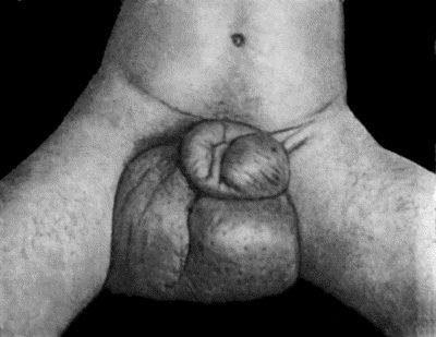
Elephantiasis from Lymphatic or Venous Obstruction.—Of this the best-known example is tropical elephantiasis (E. arabum), which is endemic in Samoa, Barbadoes, and other places. It attacks the lower extremity or the genitals in either sex (Figs. 97, 98). The disease is usually ushered in with fever, and signs of lymphangitis in the part affected. After a number of such attacks, the lymph vessels appear to become obliterated, and the skin and subcutaneous cellular tissue, being bathed in stagnant lymph—which possibly contains the products of streptococci—take on an overgrowth, which continues until the part assumes gigantic proportions. In certain cases the lymph trunks have been found to be blocked with the parent worms of the filaria Bancrofti. Cases of elephantiasis of the lower extremity are met with in this country in which there are no filarial parasites in the lymph vessels, and these present features closely resembling the tropical variety, and usually follow upon repeated attacks of lymphangitis or erysipelas.
The part affected is enormously increased in size, and causes inconvenience from its bulk and weight. In contrast to ordinary dropsy, there is no pitting on pressure, and the swelling does not disappear on elevation of the limb. The skin becomes rough and warty, and may hang down in pendulous folds. Blisters form on the surface and yield an abundant exudate of clear lymph. From neglect of cleanliness, the skin becomes the seat of eczema or even of ulceration attended with foul discharge.
Samson Handley has sought to replace the blocked lymph vessels by burying in the subcutaneous tissue of the swollen part a number of stout silk threads—lymphangioplasty. By their capillary action they drain the lymph to a healthy region above, and thus enable it to enter the circulation. It has been more successful in the face and upper limb than in the lower extremity. If the tissues are infected with pus organisms, a course of vaccines should precede the operation.
A similar type of elephantiasis may occur after extirpation of the lymph glands in the axilla or groin; in the leg in long-standing standing varix and phlebitis with chronic ulcer; in the arm as a result of extensive cancerous disease of the lymphatics in the axilla secondarily to cancer of the breast; and in extensive tuberculous disease of the lymphatics. The last-named is chiefly observed in the lower limb in young adult women, and from its following upon lupus of the toes or foot it has been called lupus elephantiasis. The tuberculous infection spreads slowly up the limb by way of the lymph vessels, and as these are obliterated the skin and cellular tissues become hypertrophied, and the surface is studded over with fungating tuberculous masses of a livid blue colour. As the more severe forms of the disease may prove dangerous to life by pyogenic complications inducing gangrene of the limb, the question of amputation may have to be considered.
Belonging to this group also is a form of congenital elephantiasis resulting from the circular constriction of a limb in utero by amniotic bands.
Elephantiasis occurring apart from lymphatic or venous obstruction is illustrated by elephantiasis nervorum, in which there is an overgrowth of the skin and cellular tissue of an extremity in association with neuro-fibromatosis of the cutaneous nerves (Fig. 89); and by elephantiasis Græcorum—a form of leprosy in which the skin of the face becomes the seat of tumour-like masses consisting of leprous nodules. It is also illustrated by elephantiasis involving the scrotum as a result of prolonged irritation by the urine in cases in which the penis has been amputated and the urine has infiltrated the scrotal tissues over a period of years.
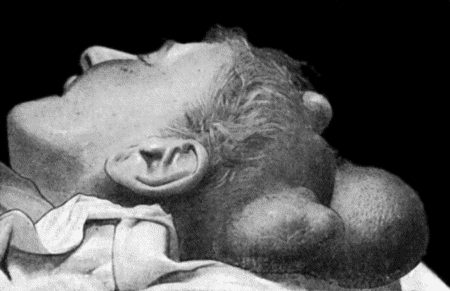
Sebaceous Cysts.—Atheromatous cysts or wens are formed in relation to the sebaceous glands and hair follicles. They are commonly met with in adults, on the scalp (Fig. 99), face, neck, back, and external genitals. Sometimes they are multiple, and they may be met with in several members of the same family. They are smooth, rounded, or discoid cysts, varying in size from a split-pea to a Tangerine orange. In consistence they are firm and elastic, or fluctuating, and are incorporated with the overlying skin, but movable on the deeper structures. The orifice of the partly blocked sebaceous follicle is sometimes visible, and the contents of the cyst can be squeezed through the opening. The wall of the cyst is composed of a connective-tissue capsule lined by stratified squamous epithelium. The contents consist of accumulated epithelial cells, and are at first dry and pearly white in appearance, but as a result of fatty degeneration they break down into a greyish-yellow pultaceous and semi-fluid material having a peculiar stale odour. It is probable that the decomposition of the contents is the result of the presence of bacteria, and that from the surgical point of view they should be regarded as infective. A sebaceous cyst may remain indefinitely without change, or may slowly increase in size, the skin over it becoming stretched and closely adherent to the cyst wall as a result of friction and pressure. The contents may ooze from the orifice of the duct and dry on the skin surface, leading to the formation of a sebaceous horn (Fig. 100). As a result of injury the cyst may undergo sudden enlargement from hæmorrhage into its interior.
Recurrent attacks of inflammation frequently occur, especially in wens of the face and scalp. Suppuration may ensue and be followed by cure of the cyst, or an offensive fungating ulcer forms which may be mistaken for epithelioma. True cancerous transformation is rare.
Wens are to be diagnosed from dermoids, from fatty tumours, and from cold abscesses. Dermoids usually appear before adult life, and as they nearly always lie beneath the fascia, the skin is movable over them. A fatty tumour is movable, and is often lobulated. The confusion with a cold abscess is most likely to occur in wens of the neck or back, and it may be impossible without the use of an exploring needle to differentiate between them.
Treatment.—The removal of wens is to be recommended while they are small and freely movable, as they are then easily shelled out after incising the overlying skin; sometimes splitting the cyst makes its removal easier. Local anæsthesia is to be preferred. It is important that none of the cyst wall be left behind. In large and adherent wens an ellipse of skin is removed along with the cyst. When inflamed, it may be impossible to dissect out the cyst, and the wall should be destroyed with carbolic acid, the resulting wound being treated by the open method.
Moles.—The term mole is applied to a pigmented, and usually hairy, patch of skin, present at or appearing shortly after birth. The colour varies from brown to black, according to the amount of melanin pigment present. The lesion consists in an overgrowth of epidermis which often presents an alveolar arrangement. Moles vary greatly in size: some are mere dots, others are as large as the palm of the hand, and occasionally a mole covers half the face. In addition to being unsightly, they bleed freely when abraded, are liable to ulcerate from friction and pressure, and occasionally become the starting-point of melanotic cancer. Rodent cancer sometimes originates in the slightly pigmented moles met with on the face. Overgrowths in relation to the cutaneous nerves, especially the plexiform neuroma, occasionally originate in pigmented moles. Soldau believes that the pigmentation and overgrowth of the epidermis in moles are associated with, and probably result from, a fibromatosis of the cutaneous nerves.
Treatment.—The quickest way to get rid of a mole is to excise it; if the edges of the gap cannot be brought together with sutures, recourse should be had to grafting. In large hairy moles of the face whose size forbids excision, radium or the X-rays should be employed. Excellent results have been obtained by refrigeration with solid carbon dioxide. In children and women with delicate skin, applications of from ten to thirty seconds suffice. In persons with coarse skin an application of one minute may be necessary, and it may have to be repeated.
Horns.—The sebaceous horn results from the accumulation of the dried contents of a wen on the surface of the skin: the sebaceous material after drying up becomes cornified, and as fresh material is added to the base the horn increases in length (Fig. 100). The wart horn grows from a warty papilloma of the skin. Cicatrix horns are formed by the heaping up of epidermis in the scars that result from burns. Nail horns are overgrown nails (keratomata of the nail bed), and are met with chiefly in the great toe of elderly bedridden patients. If an ulcer forms at the base of a horn, it may prove the starting-point of epithelioma, and for this reason, as well as for others, horns should be removed.
New Growths in the Skin and Subcutaneous Tissue.—The Angioma has been described with diseases of blood vessels. Fibroma.—Various types of fibroma occur in the skin. A soft pedunculated fibroma, about the size of a pea, is commonly met with, especially on the neck and trunk; it is usually solitary, and is easily removed with scissors. The multiple, soft fibroma known as molluscum fibrosum, which depends upon a neuro-fibromatosis of the cutaneous nerves, is described with the tumours of nerves. Hard fibromas occurring singly or in groups may be met with, especially in the skin of the buttock, and may present a local malignancy, recurring after removal like the “recurrent fibroid” of Paget. The “painful subcutaneous nodule” is a solitary fibroma related to one of the cutaneous nerves. The hard fibroma known as keloid is described with the affections of scars.
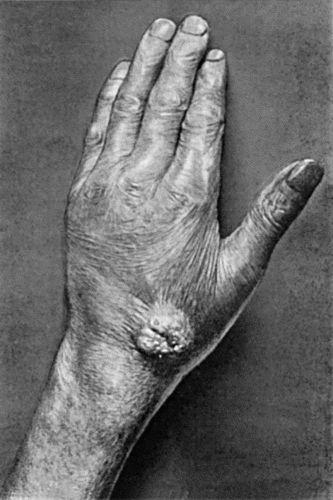
Papilloma.—The common wart or verruca is an outgrowth of the surface epidermis. It may be sessile or pedunculated hard or soft. The surface may be smooth, or fissured and foliated like a cauliflower, or it may be divided up into a number of spines. Warts are met with chiefly on the hands, and are often multiple, occurring in clusters or in successive crops. Multiple warts appear to result from some contagion, the nature of which is unknown; they sometimes occur in an epidemic form among school-children, and show a remarkable tendency to disappear spontaneously. The solitary flat-topped wart which occurs on the face of old people may, if irritated, become the seat of epithelioma. A warty growth of the epidermis is a frequent accompaniment of moles and of that variety of lupus known as lupus verrucosus.
Treatment.—In the multiple warts of children the health should be braced up by a change to the seaside. A dusting-powder, consisting of boracic acid with 5 per cent. salicylic acid, may be rubbed into the hands after washing and drying. The persistent warts of young adults should be excised after freezing with chloride of ethyl. When cutting is objected to, they may be painted night and morning with salicylic collodion, the epidermis being dehydrated with alcohol before each application.
Venereal warts occur on the genitals of either sex, and may form large cauliflower-like masses on the inner surface of the prepuce or of the labia majora. Although frequently co-existing with gonorrhœa or syphilis, they occur independently of these diseases, being probably acquired by contact with another individual suffering from warts (C. W. Cathcart). They give rise to considerable irritation and suffering, and when cleanliness is neglected there may be an offensive discharge.
In the female, the cauliflower-like masses are dissected from the labia; in the male, the prepuce is removed and the warts on the glans are snipped off with scissors. In milder cases, the warts usually disappear if the parts are kept absolutely dry and clean. A useful dusting-powder is one consisting of calamine and 5 per cent. salicylic acid; the exsiccated sulphate of iron, in the form of a powder, may be employed in cases which resist this treatment.
Adenoma.—This is a comparatively rare tumour growing from the glands of the skin. One variety, known as the “tomato tumour,” which apparently originates from the sweat glands, is met with on the scalp and face in women past middle life. These growths are often multiple; the individual tumours vary in size, and the skin, which is almost devoid of hairs, is glistening and tightly stretched over them. A similar tumour may occur on the nose. The sebaceous adenoma, which originates from the sebaceous glands, forms a projecting tumour on the face or scalp, and when the skin is irritated it may ulcerate and fungate. The treatment consists in the removal of the tumour along with the overlying skin.
The exuberant masses on the nose known as “rhinophyma,” “lipoma nasi,” or “potato nose” are of the nature of sebaceous adenoma, and are removed by shaving them off with a knife until the normal shape of the nose is restored Healing takes place with remarkable rapidity.
Cancer.—There are several types of primary cancer of the skin, the most important being squamous epithelioma, rodent cancer, and melanotic cancer.
Epithelioma occurs in a variety of forms. When originating in a small ulcer or wart-for example on the face in old people—it presents the features of a chronic indurated ulcer. A more exuberant and rapidly growing form of epithelial cancer, described by Hutchinson as the crateriform ulcer, commences on the face as a small red pimple which rapidly develops into an elevated mass shaped like a bee-hive, and breaks down in the centre. Epithelioma may develop anywhere on the body in relation to long-standing ulcers, especially that resulting from a burn or from lupus; this form usually presents an exuberant outgrowth of epidermis not unlike a cauliflower. An interesting example of epithelioma has been described by Neve of Kashmir. The natives in that province are in the habit of carrying a fire-basket suspended from the waist, which often burns the skin and causes a chronic ulcer, and many of these ulcers become the seat of epithelioma, due, in Neve's opinion, to the actual contact of the sooty pan with the skin.
The term trade epithelioma has been applied to that form met with in those who follow certain occupations, such as paraffin workers and chimney-sweeps. The most recent member of this group is the X-ray carcinoma, which is met with in those who are constantly exposed to the irritation of the X-rays; there is first a chronic dermatitis with warty overgrowth of the surface epithelium, pigmentation, and the formation of fissures and warts. The trade epithelioma varies a good deal in malignancy, but it tends to cause death in the same manner as other epitheliomas.
Epithelial cancer has also been observed in those who have taken arsenic over long periods for medicinal purposes.
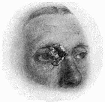
Rodent Cancer (Rodent Ulcer).—This is a cancer originating in the sweat glands or sebaceous follicles, or in the fœtal residues of cutaneous glands. The cells are small and closely packed together in alveoli or in reticulated columns; cell nests are rare. It is remarkably constant in its seat of origin, being nearly always located on the lateral aspect of the nose or in the vicinity of the lower eyelid (Fig. 102). It is rare on the trunk or limbs. It commences as a small flattened nodule in the skin, the epidermis over it being stretched and shining. The centre becomes depressed, while the margins extend in the form of an elevated ridge. Sooner or later the epidermis gives way in the centre, exposing a smooth raw surface devoid of granulations.
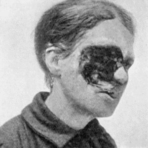
Fig. 103.—Rodent Cancer of fifteen years' duration, which has destroyed the contents of the Orbit.
(Sir Montagu Cotterill's case)
The margin, while in parts irregular, is typically represented by a well-defined “rolled” border which consists of the peripheral portion of the cancer that has not broken down. The central ulcer may temporarily heal. There is itching but little pain, and the condition progresses extremely slowly; rodent cancers which have existed for many years are frequently met with. The disease attacks and destroys every structure with which it comes in contact, such as the eyelids, the walls of the nasal cavities, and the bones of the face; hence it may produce the most hideous deformities (Fig. 103). The patient may succumb to hæmorrhage or to infective complications such as erysipelas or meningitis.
Secondary growths in the lymph glands, while not unknown, are extremely rare. We have only seen them once—in a case of rodent cancer in the groin.
Diagnosis.—Lupus is the disease most often mistaken for rodent cancer. Lupus usually begins earlier in life, it presents apple-jelly nodules, and lacks the rounded, elevated border. Syphilitic lesions progress more rapidly, and also lack the characteristic margin. The differentiation from squamous epithelioma is of considerable importance, as the latter affection spreads more rapidly, involves the lymph glands early, and is much more dangerous to life.
Treatment.—In rodent cancers of limited size—say less than one inch in diameter—free excision is the most rapid and certain method of treatment. The alternative is the application of radium or of the Röntgen rays, which, although requiring many exposures, results in cure with the minimum of disfigurement. If the cancer already covers an extensive area, or has invaded the cavity of the orbit or nose, radium or X-rays yield the best results. The effect is soon shown by the ingrowth of healthy epithelium from the surrounding skin, and at the same time the discharge is lessened. Good results are also reported from the application of carbon dioxide snow, especially when this follows upon a course of X-ray treatment.
Paget's disease of the nipple is an epithelioma occurring in women over forty years of age: a similar form of epithelioma is sometimes met with at the umbilicus or on the genitals.
Melanotic Cancer.—Under this head are included all new growths which contain an excess of melanin pigment. Many of these were formerly described as melanotic sarcoma. They nearly always originate in a pigmented mole which has been subjected to irritation. The primary growth may remain so small that its presence is not even suspected, or it may increase in size, ulcerate, and fungate. The amount of pigment varies: when small in amount the growth is brown, when abundant it is a deep black. The most remarkable feature is the rapidity with which the disease becomes disseminated along the lymphatics, the first evidence of which is an enlargement of the lymph glands. As the primary growth is often situated on the sole of the foot or in the matrix of the nail of the great toe, the femoral and inguinal glands become enlarged in succession, forming tumours much larger than the primary growth. Sometimes the dissemination involves the lymph vessels of the limb, forming a series of indurated pigmented cords and nodules (Fig. 104). Lastly, the dissemination may be universal throughout the body, and this usually occurs at a comparatively early stage. The secondary growths are deeply pigmented, being usually of a coal-black colour, and melanin pigment may be present in the urine. When recurrence takes place in or near the scar left by the operation, the cancer nodules are not necessarily pigmented.
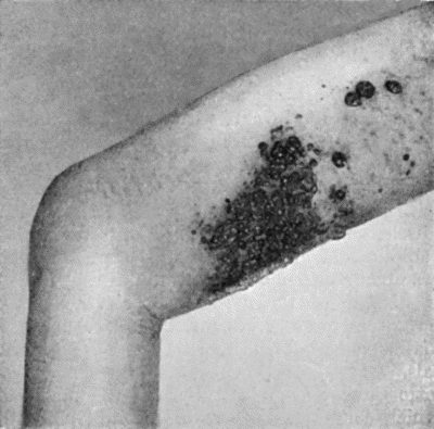
Fig. 104.—Diffuse Melanotic Cancer of Lymphatics of Skin secondary to a Growth in the Sole of the Foot.
To extirpate the disease it is necessary to excise the tumour, with a zone of healthy skin around it and a somewhat large zone of the underlying subcutaneous tissue and deep fascia. Hogarth Pringle recommends that a broad strip of subcutaneous fascia up to and including the nearest anatomical group of glands should be removed with the tumour in one continuous piece.
Secondary Cancer of the Skin.—Cancer may spread to the skin from a subjacent growth by direct continuity or by way of the lymphatics. Both of these processes are so well illustrated in cases of mammary cancer that they will be described in relation to that disease.
Sarcoma of various types is met with in the skin. The fibroma, after excision, may recur as a fibro-sarcoma. The alveolar sarcoma commences as a hard lump and increases in size until the epidermis gives way and an ulcer is formed.
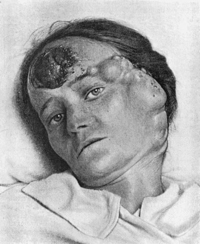
Fig. 105.—Melanotic Cancer of Forehead with Metastases in Lymph Vessels and Glands.
(Mr. D. P. D. Wilkie's case.)
A number of fresh tumours may spring up around the original growth. Sometimes the primary growth appears in the form of multiple nodules which tend to become confluent. Excision, unless performed early, is of little avail, and in any case should be followed up by exposure to radium.