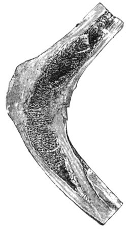
Fig. 154.—Osseous Ankylosis of Femur and Tibia in position of flexion.
Definition of Terms.—The term synovitis is applied to any reaction which affects the synovial membrane of a joint. It is usually associated with effusion of fluid, and this may be serous, sero-fibrinous, or purulent. As the term synovitis merely refers to the tissue involved, it should always be used with an adjective—such as gouty, gonorrhœal, or tuberculous—which indicates its pathological nature.
The terms hydrops, hydrarthrosis, and chronic serous synovitis are synonymous, and are employed when a serous effusion into the joint is the prominent clinical feature. Hydrops may occur apart from disease—for example, in the knee-joint from repeated sprains, or when there is a loose body in the joint—but is met with chiefly in the chronic forms of synovitis which result from gonorrhœa, tuberculosis, syphilis, arthritis deformans, or arthropathies of nerve origin.
Arthritis is the term applied when not only the synovial membrane but the articular surfaces, and it may be also the ends of the bones, are involved, and it is necessary to prefix a qualifying adjective which indicates its nature. When effusion is present, it may be serous, as in arthritis deformans, or sero-fibrinous or purulent, as in certain forms of pyogenic and tuberculous arthritis. Wasting of the muscles, especially the extensors, in the vicinity of the joint is a constant accompaniment of arthritis. On account of the involvement of the articular surfaces, arthritis is apt to be followed by ankylosis.
The term empyema is sometimes employed to indicate that the cavity of the joint contains pus. This is observed chiefly in chronic disease of pyogenic or tuberculous origin, and is usually attended with the formation of abscesses outside the joint.
Ulceration of cartilage and caries of the articular surfaces are common accompaniments of the more serious and progressive forms of joint disease, especially those of bacterial origin. The destruction of cartilage may be secondary to disease of the synovial membrane or of the subjacent bone. When the disease begins as a synovitis, the synovial membrane spreads over the articular surface, fuses with the cartilage and eats into it, causing defects or holes which are spoken of as ulcers. When the disease begins in the bone, the marrow is converted into granulation tissue, which eats into the cartilage and separates it from the bone. Following on the destruction of the cartilage, the articular surface of the bone undergoes disintegration, a condition spoken of as caries of the articular surface. The occurrence of ulceration of cartilage and of articular caries is attended with the clinical signs of fixation of the joint from involuntary muscular contraction, wasting of muscles, and starting pains. These starting pains are the result of sudden involuntary movements of the joint. They occur most frequently as the patient is dropping off to sleep; the muscles becoming relaxed, the sensitive ulcerated surfaces jar on one another, which causes sudden reflex contraction of the muscles, and the resulting movement being attended with severe pain, wakens the patient with a start. Advanced articular caries is usually associated with some abnormal attitude and with shortening of the limb. It may be possible to feel the bony surfaces grate upon one another. When all its constituent elements are damaged or destroyed, a joint is said to be disorganised. Should recovery take place, repair is usually attended with union of the opposing articular surfaces either by fibrous tissue or by bone.
Conditions of Impaired Mobility of Joints.—There are four conditions of impaired mobility in joints: rigidity, contracture, ankylosis, and locking. Rigidity is the fixation of a joint by involuntary contraction of muscles, and is of value as a sign of disease in deep-seated joints, such as the hip. It disappears under anæsthesia.
Contracture is the term applied when the fixation is due to permanent shortening of the soft parts around a joint—muscles, tendons, ligaments, fasciæ, or skin. As the structures on the flexor aspect are more liable to undergo such shortening, contracture is nearly always associated with flexion. Contracture may result from disease of the joint, or from conditions outside it—for example, disease in one of the adjacent bones, or lesions of the nerves.
Ankylosis is the term applied when impaired mobility results from changes involving the articular surfaces. It is frequently combined with contracture. Three anatomical varieties of ankylosis are recognised—(a) The fibrous, in which there are adhesions between the opposing surfaces, which may be in the form of loose isolated bands of fibrous tissue, or may bind the bones so closely together as to obliterate the cavity of the joint. The resulting stiffness, therefore, varies from a mere restriction of the normal range of movement, up to a close union of the bones which prevents movement. Fibrous ankylosis may follow upon injury, especially dislocation or fracture implicating a joint, or it may result from any form of arthritis. (b) Cartilaginous ankylosis implies the fusion of two apposed cartilaginous surfaces. It is often found between the patella and the trochlear surface of the femur in tuberculous disease of the knee. The fusion of the cartilaginous surfaces is preceded by the spreading of a vascular connective tissue, derived from the synovial membrane, over the articular cartilage. Clinically, it is associated with absolute immobility, (c) Bony ankylosis or synostosis is an osseous union between articulating surfaces (Figs. 154 and 155). It may follow upon fibrous or cartilaginous ankylosis, or may result from the fusion of two articular surfaces which have lost their cartilage and become covered with granulations. In the majority of cases it is to be regarded as a reparative process, presenting analogies with the union of fracture.
The term arthritis ossificans has been applied by Joseph Griffiths to a condition in which the articular surfaces become fused without evident cause.
The occurrence of ankylosis in a joint before the skeleton has attained maturity does not appear to impair the growth in length of the bones affected; ankylosis of the temporo-maxillary joints, however, greatly impairs the growth of the mandible. When there is arrest of growth accompanying ankylosis, it usually depends on changes in the ossifying junctions caused by the original disease.
To differentiate by manipulation between muscular fixation and ankylosis, it may be necessary to anæsthetise the patient. The nature and extent of ankylosis may be learned by skiagraphy; in osseous ankylosis the shadow of the two bones is a continuous one. In fibrous as contrasted with osseous ankylosis mobility may be elicited, although only to a limited extent; while in osseous ankylosis the joint is rigidly fixed, and attempts to move it are painless.
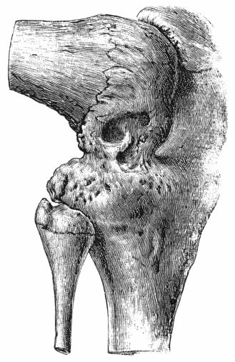
Fig. 155.—Osseous Ankylosis of Knee in the flexed position following upon Tuberculous Arthritis.
(Anatomical Museum, University of Edinburgh.)
The treatment is influenced by the nature of the original lesion, the variety of the ankylosis, and the attitude of the joint. When there is restriction of movement due to fibrous adhesions, these may be elongated or ruptured. Elongation of the adhesions may be effected by manipulations, exercises, and the use of special forms of apparatus—such as the application of weights to the limb. It may be necessary to administer an anæsthetic before rupturing strong fibrous adhesions, and this procedure must be carried out with caution, in view of such risks as fracture of the bone—which is often rarefied—or separation of an epiphysis. There is also the risk of fat embolism, and of re-starting the original disease. The giving way of adhesions may be attended with an audible crack; and the procedure is often followed by considerable pain and effusion into the joint, which necessitate rest for some days before exercises and manipulations can be resumed.
Operative treatment may be called for in cases in which the bones are closely bound to one another by fibrous or by osseous tissue.
Arthrolysis, which consists in opening the joint and dividing the fibrous adhesions, is almost inevitably followed by their reunion.
Arthroplasty.—Murphy of Chicago devised this operation for restoring movement to an ankylosed joint. It consists in transplanting between the bones a flap of fat-bearing tissue, from which a bursal cavity lined with endothelium and containing a fluid rich in mucin is ultimately formed.
Arthroplasty is most successful in ankylosis following upon injury; when the ankylosis results from some infective condition such as tuberculosis or gonorrhœa, it is liable to result in failure either because of a fresh outbreak of the infection or because the ankylosis recurs.
When arthroplasty is impracticable, and a movable joint is desired—for example at the elbow—a considerable amount of bone, and it may be also of periosteum and capsular ligament, is resected to allow of the formation of a false joint.
When bony ankylosis has occurred with the joint in an undesirable attitude—for example flexion at the hip or knee—it can sometimes be remedied by osteotomy or by a wedge-shaped resection of the bone, with or without such additional division of the contracted soft parts as will permit of the limb being placed in the attitude desired.
Bony ankylosis of the joints of a finger, whether the result of injury or disease, is difficult to remedy by any operative procedure, for while it is possible to restore mobility, the new joint is apt to be flail-like.
Locking.—A joint is said to lock when its movements are abruptly arrested by the coming together of bony outgrowths around the joint. It is best illustrated in arthritis deformans of the hip in which new bone formed round the rim of the acetabulum mechanically arrests the excursions of the head of the femur. The new bone, which limits the movements, is readily demonstrated in skiagrams; it may be removed by operative means. Locking of joints is more often met with as a result of injuries, especially in fractures occurring in the region of the elbow. In certain injuries of the semilunar menisci of the knee, also, the joint is liable to a variety of locking, which differs, however, in many respects from that described above.
Errors of Development.—These include congenital dislocations and other deformities of intra-uterine origin, such as abnormal laxity of joints, absence, displacement, or defective growth of one or other of the essential constituents of a joint. The more important of these are described along with the surgery of the Extremities.